Primary research interests
- Segmentation and tracking of structures in ultrasound images.
- Deep neural networks for medical image segmentation, object detection and classification
- Segmentation and centerline extraction of tubular structures, such as airways and blood vessels.
- Parallel and GPU processing.
My publication list can be found here.
Research projects
Interpreting ultrasound images with deep neural networks
![]()
Neural networks have recently achieved incredible results for recognizing objects such as cats, coffee cups, cars and plants in photographs. These methods are already used by companies like Facebook and Google to identify faces, recognize voice commands and even enable self-driving cars. At the Centre for Innovative Ultrasound Solutions (CIUS), we aim to use these methods to interpret ultrasound images in terms of image segmentation, object detection and classification. Ultrasound images can be challenging for humans to interpret and requires a lot of training. We believe neural networks can be used to make it easier to use ultrasound, interpret the images, extract quantitative information, and even help diagnose the patient. This may ultimately improve patient care, reduce complications and lower costs.
Efficient medical image computing and visualization on heterogeneous systems
![]()
An increasing amount of medical image data is becoming available for any given patient today. Modern image analysis techniques make it possible to extract and visualize more information from the images. The race for using the increasing amount of image data more effectively is crucial for better diagnostics and therapy in the future. Still, concurrent medical image computing and visualization of both static and dynamic real-time data is computationally expensive. In the efforts toward improving computer assisted radiology and surgery, this may entail that computational demanding research methods that have been assessed to be quantitatively better then existing methods (in terms of accuracy for example) can not be used in a routine clinical setting due to time constraints.
Most modern computer systems are heterogeneous in the sense that they consist of several different processors, such as multi-core CPUs and graphic processing units (GPUs). These processors enable parallel processing, which can accelerate many medical image computing tasks significantly. The programming of this hardware is, however, still difficult due to several factors such driver/compiler errors, the need for low-level programming and explicit memory handling.
In my PhD, I have focused on exploiting parallelism and GPUs to improve the efficiency of segmentation algorithms as well as other medical image computing and visualization algorithms. This resulted in a framework for efficient medical image computing and visualization on heterogeneous systems called FAST which I continue to work on.
RASimAs – Regional Anaesthesia Simulator and Assistant
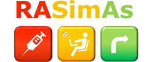
The RASimAs project (Regional Anaesthesia Simulator and Assistant) is a European research project which aims at providing a simulator to improve the training of doctors performing regional anaesthesia, as well as an assistant to lessen the cognitive burden and help performing regional anaesthesia procedures.
My role in this project is to contribute to creating an assistant for ultrasound-guided femoral nerve block.
- Streaming data directly from an ultrasound scanner to the assistant application.
- Segmentation and tracking of the important structures such as the femoral nerve, artery, fascia iliaca, fascia lata and bone.
- Registration of a 3D model to the ultrasound images.
- Visualization of the segmented ultrasound images and the 3D model.
- Visual cues to the operator guiding the correct placement of the ultrasound probe, insertion of needle, and finally injection of local anaesthetics.
Segmentation and centerline extraction of tubular structures
Blood vessels, airways and neural pathways are all examples of important tubular structures in the human body. The extraction of these structures from medical images may be useful in several medical applications. The extraction of these structures can be essential for planning and guidance of several surgical procedures such as bronchoscopy, laparoscopy, and neurosurgery. Tubular structures extracted from preoperative images can be matched to similar intraoperative structures, such as a set of positions inside the airways generated by a tracked bronchoscope, or brain vessels extracted from ultrasound.